IMMUNOHISTOCHEMISTRY
Our advanced laboratory performs Immunohistochemical (IHC) stains for the entire range of clinically relevant tissue markers.
- Over 125 antibodies
- TAT – 24-48 hours
- Technical component only or with global interpretation
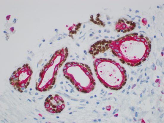
Our extensive library of antibodies includes markers for tumor differentiation, evaluation of hematologic malignancies and conditions, detection of hormone receptors, and identification of viruses, among others. Our specialized technologists are extensively trained and highly proficient with the most advanced immunohistochemical methods. New antibodies are added to our inventory on a regular basis as new immunohistochemical stains are developed.
LISTING OF IHC STAINS
(click on stains for description or download IHC Library here)
-
Actin
STAIN NAME
Actin
ANTIBODY
(HHF35) Muscle Specific
DESCRIPTION
Helps to identify smooth muscle tumor and myoepithelial cells
-
AE1/AE3 (Cytokeratin Cocktail)
STAIN NAME
AE1/AE3 (Cytokeratin Cocktail)
ANTIBODY
Cytokeratin AE1/AE3
DESCRIPTION
Can be used to differentiate epithelial from non-epithelial tumors
-
AFP (Alpha-1-Fetoprotein)
STAIN NAME
AFP (Alpha-1-Fetoprotein)
ANTIBODY
Polyclonal Anti-human
DESCRIPTION
Helps to identify hepatocellular carcinomas, gonadal and extragonadal yolk sac tumors and mixed germ cell tumors
-
ALK-1
STAIN NAME
ALK-1
ANTIBODY
ALK-1
DESCRIPTION
Helps to identify anaplastic large cell lymphomas
-
AMACR (P504S)
STAIN NAME
AMACR (P504S)
ANTIBODY
13H4
DESCRIPTION
Aids in the classification of premalignant high-grade prostatic intraepithelial neoplasia and prostate adenocarcinoma
-
Androgen Receptor
STAIN NAME
Androgen Receptor
ANTIBODY
Monoclonal Mouse Androgen Receptor
DESCRIPTION
In skin, positive staining has been observed in cells of hair follicles, sweat glands and squamous cells of the epidermis. In adenocarcinomas of the prostate androgen receptor has been demonstrated by IHC to be present in the nuclei of most neoplastic cell with neuroendocrine differentiation. Strong to moderate expressions in cervical, ovarian cells and placenta.
-
APP (Amyloid Precursor Protein)
STAIN NAME
APP (Amyloid Precursor Protein)
ANTIBODY
APP
DESCRIPTION
Helps with the identification of Alzheimer’s Disease
-
Arginase-1
STAIN NAME
Arginase-1
ANTIBODY
Rabbit Monoclonal
DESCRIPTION
Helps to distinguish hepatocellular carcinoma from a metastatic tumor to the liver
-
BCL1 (Cyclin D1)
STAIN NAME
BCL1 (Cyclin D1)
ANTIBODY
SP4
DESCRIPTION
Useful in identification of mantle cell lymphomas
-
BCL2
STAIN NAME
BCL2
ANTIBODY
Clone 124
DESCRIPTION
Useful in identification of follicular lymphomas and disease progression
-
BCL6
STAIN NAME
BCL6
ANTIBODY
PG-B6p
DESCRIPTION
Useful in identification of follicular lymphoma, DLBCL and Burkitt Lymphoma
-
BER-EP4 (Epithelial Antigen)
STAIN NAME
BER-EP4 (Epithelial Antigen)
ANTIBODY
BER-EP4
DESCRIPTION
Helps to differentiate carcinomas from mesotheliomas
-
Beta-Catenin
STAIN NAME
Beta-Catenin
ANTIBODY
Beta-Catenin
DESCRIPTION
Used to distinguish deep fibromatosis from other benign and malignant fibroblastic and
myofibroblastic lesion, primary adenocarcinoma of the bladder and secondary involvement by
colorectal adenocarcinoma, distinguish hepatocellular carcinoma from focal nodular
hyperplasia, and aid in the diagnosis of solid pseudopapillary neoplasms -
CA125
STAIN NAME
CA125
ANTIBODY
M11
DESCRIPTION
Useful in the idenification of ovarian carcinomas
-
CA19-9
STAIN NAME
CA19-9
ANTIBODY
116-NS-19-9
DESCRIPTION
Useful in the identification of GI carcinoma
-
Calponin
STAIN NAME
Calponin
ANTIBODY
Clone CALP
DESCRIPTION
Myoepithelial marker, in situ vs. infiltrating breast carcinoma
-
Calretinin
STAIN NAME
Calretinin
ANTIBODY
DAK Calret 1
DESCRIPTION
Most specific and reproducible positive marker of epithelial mesothelioma
-
CAM 5.2
STAIN NAME
CAM 5.2
ANTIBODY
CAM 5.2
DESCRIPTION
This antibody is used to aid in the identification of tumors of epithelial origin and in
distinguishing carcinomas from other malignant tumors of non-epithelial origin -
Carbonic Anhydrase (CA IX)
STAIN NAME
Carbonic Anhydrase (CA IX)
ANTIBODY
M75
DESCRIPTION
Helpful in clear cell renal cell carcinoma, and strong diffuse-to multifocal staining in large
majority of urothelial carcinomas, as opposed to extremely weak and focal staining in collecting
duct carcinoma -
CD10
STAIN NAME
CD10
ANTIBODY
56C6
DESCRIPTION
Expressed in early lymphoid progenitor and normal germinal center cells
-
CD117
STAIN NAME
CD117
ANTIBODY
c-kit (polyclonal)
DESCRIPTION
Aid in the differentiation of GIST, management of (Gleevec®) therapy
-
CD138
STAIN NAME
CD138
ANTIBODY
MI 15
DESCRIPTION
Positive in B-cell precursors, plasma cells, myeloma
-
CD15
STAIN NAME
CD15
ANTIBODY
Carb-3
DESCRIPTION
Useful in the identification of Reed-Sternberg cells and Hodgkin lyphomas
-
CD163
STAIN NAME
CD163
ANTIBODY
MRQ-26
DESCRIPTION
Anti-CD163 labels cells of monocytic-macrophage lineage, with expression in bone marrow and histiocytic neoplasms.
-
CD1a
STAIN NAME
CD1a
ANTIBODY
Clone: 010
DESCRIPTION
Expressed on cortical thymocytes, Langerhans cells, and dendritic cells
-
CD2
STAIN NAME
CD2
ANTIBODY
AB75
DESCRIPTION
Labels CD2-expressing normal T cells and related neoplasms. Results aid in the classification of precursor T-cell lymphoma, peripheral
T-cell lymphoma, and anaplastic large cell lymphoma. -
CD20
STAIN NAME
CD20
ANTIBODY
Pan B-Cell
DESCRIPTION
Labels normal and neoplastic B-cells and if absent from plasma cells
-
CD21
STAIN NAME
CD21
ANTIBODY
IF8
DESCRIPTION
Expressed on mature B-cells, folllicular dendritic cells (FDC)
-
CD23
STAIN NAME
CD23
ANTIBODY
DAK-CD23
DESCRIPTION
Useful in distinguishing B-cells CLL/SLL from other entities
-
CD25
STAIN NAME
CD25
ANTIBODY
4C9
DESCRIPTION
CD25 has been detected in various hematological malignancies including adult T-cell leukemia/lymphoma, and hairy cell leukemia
-
CD3
STAIN NAME
CD3
ANTIBODY
Pan T-cell
DESCRIPTION
Labels normal and neoplastic T-cells
-
CD3/CD20
STAIN NAME
CD3/CD20
ANTIBODY
CD3/CD20 double stain
DESCRIPTION
Differentiates between B and T cells
-
CD30
STAIN NAME
CD30
ANTIBODY
BER-H2
DESCRIPTION
Labels Reed-Sternberg cells, ALCL, T-cell lymphomas, germ cell tumors
-
CD31
STAIN NAME
CD31
ANTIBODY
JC7041
DESCRIPTION
Endothelial cells and prognostic marker for tumor angiogenesis
-
CD34
STAIN NAME
CD34
ANTIBODY
Hematopoietic Progenitor QBEnd 10
DESCRIPTION
Expressed on lymphoid, myeloid, endothelial GIST
-
CD4/CD8
STAIN NAME
CD4/CD8
ANTIBODY
MT310 & DK25
DESCRIPTION
Used to differentiate cutaneous T-cell lymphomas, including mycoses fungoides
-
CD43
STAIN NAME
CD43
ANTIBODY
T-cell DF-T1
DESCRIPTION
Diagnosis of T-cell lymphoma and a subset of B-cell lymphoma
-
CD44
STAIN NAME
CD44
ANTIBODY
DF1485
DESCRIPTION
Transitional carcinoma-in-situ vs. non-neoplatic changes in the urothelium
-
CD45 (LCA)
STAIN NAME
CD45 (LCA)
ANTIBODY
2B11+PD7/26
DESCRIPTION
Labels all hematopoietic cells
-
CD5
STAIN NAME
CD5
ANTIBODY
SP19
DESCRIPTION
Useful in the identification of B-cells, MCL, HCL, CLL/SLL, T-cells, thymic carcinomas
-
CD56
STAIN NAME
CD56
ANTIBODY
123C3
DESCRIPTION
Neuroedrocrine marker, NK cells
-
CD68
STAIN NAME
CD68
ANTIBODY
KP1
DESCRIPTION
Labels macrophages myeloid precursors, preipheral blood granulocytes
-
CD7
STAIN NAME
CD7
ANTIBODY
Clone: CBC.37
DESCRIPTION
Lables immature and mature T-lymphocytes and T-cell Leukemia
-
CD79a
STAIN NAME
CD79a
ANTIBODY
Pan B-cell JBC1171
DESCRIPTION
Acute leukemias, B-cell lymphomas, and in some myelomas
-
CD99
STAIN NAME
CD99
ANTIBODY
12E71
DESCRIPTION
Expressed by Ewing’s sarcoma cells and lymphoblastic leukemia/lymphoma
-
CDX2
STAIN NAME
CDX2
ANTIBODY
DAK-CDX2
DESCRIPTION
Expressed in primary and metastatic colorectal and gastric carcinomas
-
CEA
STAIN NAME
CEA
ANTIBODY
Carcinoembyronic Antigen
DESCRIPTION
Useful in identifying the origin of various metastatic adenocarcinomas
-
Chromogranin A
STAIN NAME
Chromogranin A
ANTIBODY
Chromogranin A (polyclonal)
DESCRIPTION
Useful in identification of neuroendocrine tumors
-
CK19
STAIN NAME
CK19
ANTIBODY
RCK 108
DESCRIPTION
Useful in the distinction of liver metastasis from hepatocellular carcinomas
-
CK20
STAIN NAME
CK20
ANTIBODY
Ks20.8
DESCRIPTION
Found in colon, ovarian, stomach, bile, pancreas and Merkel Cells
-
CK5/6
STAIN NAME
CK5/6
ANTIBODY
D5 / 16B4
DESCRIPTION
Labels mesotheliomas, transitional and squamous cell carcinomas
-
CK7
STAIN NAME
CK7
ANTIBODY
OV-TL 12/30
DESCRIPTION
Identify positive lung and breast and negative colon and prostate cells
-
CK903
STAIN NAME
CK903
ANTIBODY
34bE12
DESCRIPTION
Can be used for the identification of basal cell layer in prostate tissue
-
CMV
STAIN NAME
CMV
ANTIBODY
DDG9 + CCH2
DESCRIPTION
Useful in identification of CMV infection
-
Collagen IV
STAIN NAME
Collagen IV
ANTIBODY
CIV 22
DESCRIPTION
Useful in detecting the loss of parts of basement membrane in carcinomas
-
D2-40
STAIN NAME
D2-40
ANTIBODY
D2-40
DESCRIPTION
Lymphatic invasion of primary tumors; seminomas and mesotheliomas
-
Desmin
STAIN NAME
Desmin
ANTIBODY
D33
DESCRIPTION
Reacts with leiomyosarcomas and cells such as rhabdomyosarcomas
-
Dog-1
STAIN NAME
Dog-1
ANTIBODY
DOG1.1
DESCRIPTION
Used as a very sensitive and specific marker for GIST
-
EBER-ISH
STAIN NAME
EBER-ISH
ANTIBODY
ISH
DESCRIPTION
Used in the detection of the Epstein Barr Virus (EBV) RNA
-
EBV
STAIN NAME
EBV
ANTIBODY
CS.1-4
DESCRIPTION
Used in the detection of the Epstein Barr Virus (EBV)-positive lymphoblastoid cell lines and
EBV infected B-cell immunoblasts in infectious mononucleosis -
E-cadherin
STAIN NAME
E-cadherin
ANTIBODY
NCH-38
DESCRIPTION
Useful in the differentiation of ductal and lobular carcinomas of the breast
-
EMA
STAIN NAME
EMA
ANTIBODY
E29
DESCRIPTION
Stains normal and neoplastic cells from various carcinomas
-
ER
STAIN NAME
ER
ANTIBODY
EP1
DESCRIPTION
Predicts response of breast cancer to anti-hormonal therapy, such as tamoxifen
-
ERG
STAIN NAME
ERG
ANTIBODY
EP111
DESCRIPTION
Used to identify prostate carcinoma; also as an endothelial cell marker
-
Factor VIII (vWF:Ag)
STAIN NAME
Factor VIII (vWF:Ag)
ANTIBODY
polyclonal anti-VWF
DESCRIPTION
Diagnosis of vascular neoplasms
-
Factor XIIIa
STAIN NAME
Factor XIIIa
ANTIBODY
E980.1
DESCRIPTION
Differentiate between dermatofibroma and dermatofibrosarcoma
-
GATA3
STAIN NAME
GATA3
ANTIBODY
GATA3
DESCRIPTION
Used in the IHC workup panel for unknown primary carcinomas, when carcinomas of the breast or urinary bladder are a possibility
-
GCDFP-15
STAIN NAME
GCDFP-15
ANTIBODY
Gross Cystic Disease Fluid Protein
DESCRIPTION
Breast carinoma, salivary duct carcinoma and apocrine epithelia
-
GFAP
STAIN NAME
GFAP
ANTIBODY
polyclonal
DESCRIPTION
Differentiate the primary gliomas from metastatic lesions in the brain
-
Glypican-3
STAIN NAME
Glypican-3
ANTIBODY
Glypican-3
DESCRIPTION
Differentiate hepatocellular carcinomas from normal liver tissue and benign hepatic lesion
-
HBME-1
STAIN NAME
HBME-1
ANTIBODY
(clone HBME-1)
DESCRIPTION
Reacts with mesothelial cells, mesotheliomas and some (20-30%) carcinomas
-
h-Caldesmon
STAIN NAME
h-Caldesmon
ANTIBODY
h-CD
DESCRIPTION
Helps identify smooth muscle tumors
-
HCG(chorionic gonadotropin)
STAIN NAME
HCG(chorionic gonadotropin)
ANTIBODY
Polyclonal
DESCRIPTION
Identification of gestational trophoblastic tumors derived from the placenta
-
Helicobacter pylori
STAIN NAME
Helicobacter pylori
ANTIBODY
B0471
DESCRIPTION
Reacts with H. pylori on the surface of epithelial cells of stomach biopsies
-
HEP-PAR1 (Hepatocyte)
STAIN NAME
HEP-PAR1 (Hepatocyte)
ANTIBODY
OCH1E5
DESCRIPTION
Differentiate hepatocellular carinomas from adenocarinomas
-
HER2 DISH
STAIN NAME
HER2 DISH
ANTIBODY
DNA Probe
DESCRIPTION
HER2 protien over-expression and aids in assessment of patients being considered for Herceptin treatment
-
HER2 IHC
STAIN NAME
HER2 IHC
ANTIBODY
Herceptest
DESCRIPTION
HER2 protien over-expression, patients may benefit from trastuzumab (Herceptin) therapy
-
Hgb A
STAIN NAME
Hgb A
ANTIBODY
Polyclonal
DESCRIPTION
Useful in the detection of red blood cell precursors
-
HHV-8
STAIN NAME
HHV-8
ANTIBODY
13B10
DESCRIPTION
Identification of the latent nuclear protein of the human herpes virus 8 in Kaposi’s Sarcoma,
multicentric Castleman’s disease, and primary effusion lymphoma -
HMB 45
STAIN NAME
HMB 45
ANTIBODY
HMB45
DESCRIPTION
Expressed on melanomas and tumors of melanocytic differentiation
-
HSV1
STAIN NAME
HSV1
ANTIBODY
Polyclonal
DESCRIPTION
Detection of HSV 1 in human cellular material
-
HSV2
STAIN NAME
HSV2
ANTIBODY
Polyclonal
DESCRIPTION
Detection of HSV 2 in human cellular material
-
Inhibin
STAIN NAME
Inhibin
ANTIBODY
R1
DESCRIPTION
Differentiation between adrenocortical tumors and renal cell carcinoma
-
Kappa- ISH
STAIN NAME
Kappa- ISH
ANTIBODY
DNA Probe
DESCRIPTION
Reacts with immunoglobulin Kappa-light chain mRNA
-
Kappa Light chain
STAIN NAME
Kappa Light chain
ANTIBODY
Polyclonal
DESCRIPTION
Reacts with immunoglobulin Kappa-light chain
-
Kappa/Lambda Double stain
STAIN NAME
Kappa/Lambda Double stain
ANTIBODY
KDB-1 + N/A
DESCRIPTION
Differentiation between Kappa and Lambda by IHC
-
Kappa/Lambda Double stain – ISH
STAIN NAME
Kappa/Lambda Double stain – ISH
ANTIBODY
DNA Probe
DESCRIPTION
Differentiation between Kappa and Lambda by mRNA.
-
Ki67
STAIN NAME
Ki67
ANTIBODY
MIB-1
DESCRIPTION
Labels proliferating cells in late G1-, S-, M-, and G2 phases of the cell cycle
-
Lambda Light chain
STAIN NAME
Lambda Light chain
ANTIBODY
Polyclonal
DESCRIPTION
Reacts with immunoglobulin Lambda-light chain
-
Lambda- ISH
STAIN NAME
Lambda- ISH
ANTIBODY
DNA Probe
DESCRIPTION
Reacts with immunoglobulin Lambda-light chain mRNA
-
Lysozyme (Muramidase)
STAIN NAME
Lysozyme (Muramidase)
ANTIBODY
Polyclonal
DESCRIPTION
Helps to identify myeloid cells, monocytes, histiocytes, and poorly differentiated leukemic
monoblasts. Lysozyme is also expressed in myeloproliferative disorders. -
Mammaglobin
STAIN NAME
Mammaglobin
ANTIBODY
304-1A5
DESCRIPTION
Useful in the identification of breast carcinoma
-
Melan A
STAIN NAME
Melan A
ANTIBODY
A103
DESCRIPTION
Melanomas, adrenocortical carincomas and sex cord tumors of the gonads
-
Microinvasion cocktail (CK5/6, p63, CK8/18)
STAIN NAME
Microinvasion cocktail (CK5/6, p63, CK8/18)
ANTIBODY
CK 5/6 & p63 with CK8/18
DESCRIPTION
Distinguishing DCIS from microinvasive breast carcinoma
-
MITF
STAIN NAME
MITF
ANTIBODY
MITF
DESCRIPTION
Aids in the classification of melanoma
-
MMR IHC Panel for MSI (MLH1, MSH2, MSH6, PMS2)
STAIN NAME
MMR IHC Panel for MSI (MLH1, MSH2, MSH6, PMS2)
ANTIBODY
ES05,F811,EP49, EP51
DESCRIPTION
Absence correlates with a mismatch repair gene defect in the respective gene
-
MOC31
STAIN NAME
MOC31
ANTIBODY
MOC 31
DESCRIPTION
Helps to differentiate carcinomas from mesotheliomas
-
MPO
STAIN NAME
MPO
ANTIBODY
Myeloperoxidase
DESCRIPTION
Useful in differentiating between myeloid and lymphoid leukemias
-
MUM 1
STAIN NAME
MUM 1
ANTIBODY
MUM1p
DESCRIPTION
Associated with poor prognosis of patients with diffuse large B-cell Lymphoma
-
Myo D1
STAIN NAME
Myo D1
ANTIBODY
(clone: 5.8A)
DESCRIPTION
Stains majority of rhabdomyosarcomas of various histological subtypes
-
Napsin A
STAIN NAME
Napsin A
ANTIBODY
Polyclonal
DESCRIPTION
Used in the identification of pulmonary adenocarcinomas
-
NSE
STAIN NAME
NSE
ANTIBODY
BBS/NC/VI-H14
DESCRIPTION
Neuroendocrine tumors
-
Oct-4
STAIN NAME
Oct-4
ANTIBODY
MRQ-10
DESCRIPTION
High sensitivity and specificity in seminoma/dysgerminoma, embryonal carcinoma and a germ
component of gonadoblastoma -
P16
STAIN NAME
P16
ANTIBODY
INK4a
DESCRIPTION
Overexpression reported for some cancer entities
-
P40
STAIN NAME
P40
ANTIBODY
BC28
DESCRIPTION
Used as an ancillary tool for a diagnosis of pulmonary squamous cell carcinoma
-
P53
STAIN NAME
P53
ANTIBODY
DO-7
DESCRIPTION
Overexpression associated with high proliferative rates
-
P57
STAIN NAME
P57
ANTIBODY
Kp10
DESCRIPTION
Useful in the identification of complete moles versus partial mole or hydropic abortus
-
P63
STAIN NAME
P63
ANTIBODY
BC4A4
DESCRIPTION
Marker of myoepithelial and basal cells
-
Parathyroid Hormone
STAIN NAME
Parathyroid Hormone
ANTIBODY
MRQ-31
DESCRIPTION
Anti-PTH antibody is useful to distinguish parathyroid hyperplasia/neoplasms from thyroid and metastatic neoplasms
-
PAX5
STAIN NAME
PAX5
ANTIBODY
BC/24
DESCRIPTION
Labels normal and neoplastic B-cells
-
PAX8
STAIN NAME
PAX8
ANTIBODY
Polyclonal
DESCRIPTION
Identifies clear cell, chromophobe and papillary renal carcinoma, negative in urothelium; stains tumors of Mullerian origin
-
PD-L1 22C3
STAIN NAME
PD-L1 22C3
ANTIBODY
22C3
DESCRIPTION
Used in non-small cell cancer of lung to see if a patient is eligible for Keytruda®
-
PD-L1 28-8
STAIN NAME
PD-L1 28-8
ANTIBODY
28-8
DESCRIPTION
Used in non-squamous, non-small cell cancer of lung to see if a patient is eligible for Opdivo®.
-
Phosphohistone H3 (PHH3)
STAIN NAME
Phosphohistone H3 (PHH3)
ANTIBODY
PHH3
DESCRIPTION
PHH3 is a core histone protein, one of the major proteins that reach a maximum for chromatin
condensation during mitosis; it serves as a mitotic marker to separate mitotic figures from
apoptotic bodies and karyorrhectic debris (due to their lack of phosphorylations) -
PIN4 Triple Stain (CK5/14, p63, P504S)
STAIN NAME
PIN4 Triple Stain (CK5/14, p63, P504S)
ANTIBODY
P504S (AMACR) & P63 & HMW CK (XM26 + LL002 + BC4A4 + N/A)
DESCRIPTION
Helps with diagnosing prostatic intraepithelial neoplasia
-
PLAP
STAIN NAME
PLAP
ANTIBODY
8A9
DESCRIPTION
Useful in the identification of seminoma and embryonal carcinomas
-
PR
STAIN NAME
PR
ANTIBODY
PgR 636
DESCRIPTION
Predicts response of breast cancer to anti-hormonal therapy, such as tamoxifen
-
PRAME
STAIN NAME
PRAME
ANTIBODY
EPR20330
DESCRIPTION
Preferentially expressed antigen in melanoma. An autosomal cancer-testis antigen (CTA) gene.
-
PSA
STAIN NAME
PSA
ANTIBODY
Polyclonal
DESCRIPTION
Useful in the identification of metastasis or prostatic origin
-
RCC
STAIN NAME
RCC
ANTIBODY
SPM314
DESCRIPTION
Expressed in renal cell carcinomas and normal kidney tissue
-
S100
STAIN NAME
S100
ANTIBODY
Polyclonal
DESCRIPTION
Schwannomas, ependymomas, astrogliomas, benign/malignant melanomas
-
SMM-HC
STAIN NAME
SMM-HC
ANTIBODY
SMMS-1
DESCRIPTION
Labels smooth muscle and myoepthithelial cells, and sclerosing breast lesions
-
SOX-10
STAIN NAME
SOX-10
ANTIBODY
BC34
DESCRIPTION
Sensitive marker for neoplasms of neural crest origin (eg Melanoma) and does not label
carcinomas -
SOX-11
STAIN NAME
SOX-11
ANTIBODY
MRQ-58
DESCRIPTION
Specific marker for cyclin D1-positive and cyclin D1-negative MCL
-
Synaptophysin
STAIN NAME
Synaptophysin
ANTIBODY
DAK-SYNAP
DESCRIPTION
Identification of normal neuroendocrine cells and neuroendocrine neoplasms
-
TdT
STAIN NAME
TdT
ANTIBODY
Polyclonal
DESCRIPTION
Helpful for the diagnosis of acute lymphoblastic lymphoma/leukemias
-
Thrombomodulin
STAIN NAME
Thrombomodulin
ANTIBODY
1009
DESCRIPTION
Labels most mesotheliomas with a membranous staining pattern
-
Thyroglobulin
STAIN NAME
Thyroglobulin
ANTIBODY
Polyclonal
DESCRIPTION
Identification of thyroid carcinomas of the papillary and follicular types
-
TTF-1
STAIN NAME
TTF-1
ANTIBODY
8G7G3/1
DESCRIPTION
Reactive with normal and neoplastic thyroid and lung adenocarcinomas
-
TTF-1/Napsin A (Double stain) lung adenocarcinoma
STAIN NAME
TTF-1/Napsin A (Double stain) lung adenocarcinoma
ANTIBODY
8G7G3/1 + NA
DESCRIPTION
A useful marker for identifying lung adenocarcinoma
-
Vimentin
STAIN NAME
Vimentin
ANTIBODY
V9
DESCRIPTION
Mesenchymal cells, lymphocytes, sarcomas, melanomas
-
WT1
STAIN NAME
WT1
ANTIBODY
6F-H2
DESCRIPTION
Mesothelioma, ovarian serous tumors, renal cell carcinoma, and Wilm’s tumor