A Guide to Immunocytochemistry and its Applications in Cytopathology
By Ing Wei Khor
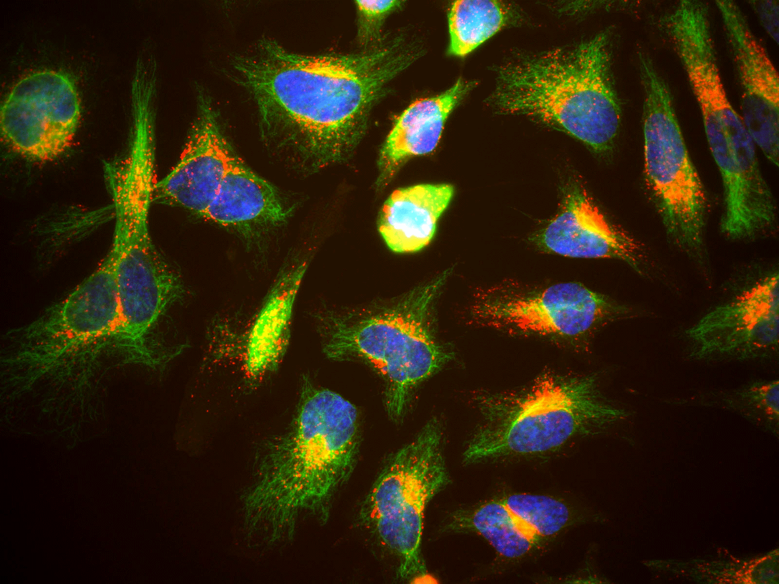
Immunocytochemistry is a method for detecting a specific protein in cells by using an antibody that binds to the protein. This method is often used by pathologists to identify cancer cells and aid in cancer diagnosis, in a subfield of pathology called cytopathology. Cancer cells produce specific proteins which normal cells do not. In cytopathology, pathologists use immunocytochemistry as one method to detect these cancer-specific proteins and thus determine that cancer cells are present. It is an indispensable tool in anatomic pathology laboratories, where pathologists analyze cells obtained via biopsies or smears to aid in the diagnosis and management of disease.
How Does Immunocytochemistry Work?
Cell Adherence
The cells are placed on a glass slide or a cell culture plate with a glass bottom. The cells are allowed to adhere or stick to the glass, which may take an hour to more than a day, depending on the type of cell. Another option is to grow the cells and perform immunocytochemistry on the same glass surface. Cells are cultured in the cell culture medium (a special solution containing proteins) in a special chamber attached to a glass slide. Once enough cells are grown and adhere to the slide, the chamber containing the medium is discarded.
Cell Fixation and Permeabilization
The cells on the glass slide or plate are fixed, usually with a formalin solution, which kills the cells. The fixed cells are then permeabilized using a detergent solution, to enable the antibody molecules to get into the cells and bind to the proteins inside the cell, along with proteins on the cell surface.
Cell Staining
The staining step involves applying antibodies that bind to and “stain” or label the protein of interest. A common immunocytochemistry method uses antibodies with a fluorescent dye attached to them. After allowing some time (1 h to several hours at room temperature or refrigerated overnight) for the antibodies to bind, the excess unbound antibody is washed off. Very often, using only one type of antibody (called the primary antibody) is insufficient for detecting proteins in cells because there are too few of these proteins. Therefore, pathologists and pathology laboratories usually apply another antibody (the secondary antibody). In this method, the secondary antibody is labeled with a fluorescent dye instead of the primary antibody. This secondary antibody binds to the primary antibody. Since several secondary antibody molecules bind to one primary antibody molecule, this system amplifies the fluorescent signal produced by binding it to the protein of interest.
Another staining procedure uses an antibody attached to an enzyme, such as horseradish peroxidase. When the enzyme-linked antibody recognizes and binds to the specific protein, the peroxidase enzyme changes the substance 3,3′-diaminobenzidine (DAB) into a brown product, which is detectable.
Detection of Specific Proteins
To determine whether a particular protein is present, pathologists can detect fluorescent antibodies specific for that protein using a fluorescence microscope. Or, if enzyme-linked antibodies specific for the protein are applied, pathologists can use a light microscope to detect the product from the reaction of the enzyme-linked antibody and DAB. They can also measure the amount of fluorescence to get an idea of how much of the protein is present.
Applications of Immunocytochemistry in Cytopathology
Immunocytochemistry helps pathologists to identify specific proteins in cells such as cancer cells, which can in turn aid in diagnosing cancer. The method is useful for cancer screening, for example, screening for cervical cancer by analyzing Pap smears and confirming a cancer diagnosis in patients who have physical or laboratory signs of cancer.

