Whole Slide Imaging: Facts, Benefits and Barriers Within Pathology
By Ing Wei Khor
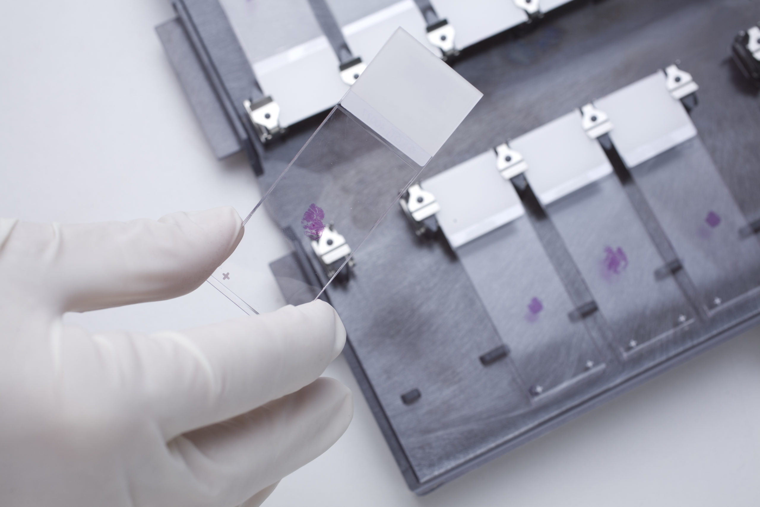
Whole slide imaging is a relatively new technique that is becoming increasingly important in anatomic and clinical pathology laboratories, where pathologists analyze tissues and organs to help diagnose and manage disease. The method involves digitally scanning a tissue slide so that pathologists can analyze the digital images, share the images quickly and conveniently with other clinicians around the world, and compare their whole slide images with image banks to help them make an accurate diagnosis in a timely manner.
How is Whole Slide Imaging Performed?
In whole slide imaging, a specialized microscope is used to scan a slide in an automated way and create a digital, high-resolution image of the entire slide by piecing many separate images together. This digital image may require specific software for viewing and analysis.
How is Whole Slide Imaging Different from Examining Slides Under a Microscope?
Opinions in the field of anatomic pathology vary about the differences between whole slide imaging and manual viewing of tissue slides under a light microscope. Based on guidelines such as those put forth by the College of American Pathologists for validating whole slide imaging, many studies have shown that the diagnosis established using whole slide imaging showed a high rate of agreement with diagnoses obtained using light microscopy. At least one whole slide imaging platform has been approved for diagnostic use in the U.S., with formalin-fixed, paraffin-embedded tissue sections.
What are the Benefits of Whole Slide Imaging?
Whole slide imaging enables many tissue slides to be scanned in a short time (high-throughput scanning), which may reduce the time spent on viewing and searching for an optimal area of a tissue slide under the microscope. The method also makes it possible for pathologists to consult with their colleagues at hospitals in the U.S. and other countries about difficult or ambiguous tissue specimens, or as part of collaborations in clinical trials or research studies. Another benefit of whole slide imaging is that the images are easily stored in secure image banks, allowing convenient access by pathologists who wish to compare their images with those obtained in previous clinical evaluations or research studies.
Why Aren’t All Pathology Laboratories Using This Method?
It’s relatively new, thus some pathologists and pathology laboratories may be reluctant to adopt it until they learn more about it and hear about other laboratories’ experiences with and feedback about the method. Whole slide imaging also requires specialized equipment, such as a microscope equipped with a whole slide scanner, as well as image viewing software. Pathologists need to be trained to use the specialized equipment and software, and to view and analyze the digital images. The need for such an investment in equipment and training could be a barrier for some laboratories. However, despite these barriers, many pathology laboratories are starting to adopt whole slide imaging because of the significant advantages it affords in terms of convenience, sustainability and advancement of pathology practice and research.